Auditory Cues and Ecolocation
Sound
- Intensity
- Frequency
- Direction
Prey detection
Object localization
Owls
Vision & hearing
Home ranges
If target detected:
- Orient head
- Scrutinize source
- Capture
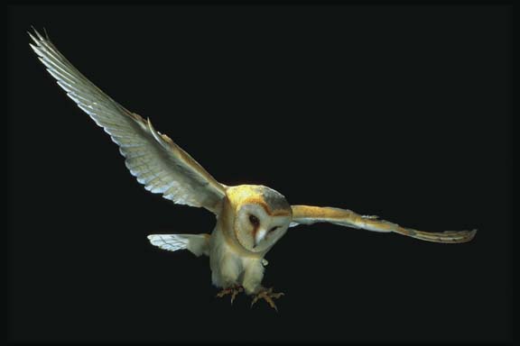
The hunting technique of an owl, drawn from photographs of Tengmalm's owl, Aegolius funereus, in its natural habitat. (a) The owl about to strike the prey with its talons, after flying down from an observation perch. (b) The owl on its perch immediately before striking, with a diagram showing the erros involved in localising prey by hearing. The prey (o) is observed at a shallow angle (alpha), with the result that a given angle of error converts into a greater distance along the ground for a vertical (elevation) error than for a horizontal (azimuth) error. (Modified after Norberg, 1970. 1977). (From: Young, 1989, p. 189.)
Horizontal (azimuth) and vertical (elevation)
Can orient in full darkness by sound
Greatest accuracy 6 to 9 kHz
Experimental apparatus
Appratus used to measure accuracy of owls in localization of sounds from different positions in space. (From: Young, 1989, p. 191.)
Do not localize through approximations
Relatively low error rate
Localization of sound accuracy as a function of sound position in space, showing mean degree of error in localization of sound target in horizontal (left) and vertical plane (right) for an individual owl. (From: Young, 1989, p. 191.)
Intensity difference between ears
Intensity --> elevation information
Ear plug experiments
- Left --> above & right of target
- Right --> below and left of target
Facial asymmetries
- Left ear above midpoint of eyes
- Right ear below midpoint of eyes
- Facial ruff
Barn owl's ability to localize sounds in elevation. (a) Plot of auditory space in front of owl in degrees of azimuth (L and R) and elevation (+ and -). (b) Facial ruff of the barn owl. (From Young, 1989, p. 192.)
Ruff removed
- Can't orient in vertical plane
- Azimuth unaffected
Time differences between ears
Time difference --> azimuth direction
Earphones
- Same intensity, different times
- Turns to side that receives sound 1st
- Rotation has positive correlation with time difference

Owl Brain Structure for Audition
Optic tectum
Neuronal map of auditory space in the midbrain of the barn owl. The bottom element of the figure depicts the receptive fields (bold rectangles) of 10 neurons recorded in three separate electrode penetrations. (From: Young, 1989, 195.)
Inner region interneurons
- Particular frequency
- Arranged tonotopically
Outer region interneurons
- Particular spatial location
- Limited-field neurons
- Insensitive to sound changes
Limited-field neurons
- Neuronal map of auditory space
- 30 degrees in front

Receptive fields of auditory interneurons in midbrain of the barn owl. (From: Young, 1989, p. 194.)
Sonar
SOund NAvigtation Ranging
Detect underwater objects
Transmitter
Receiver
Time for echo to return --> depth
--Fig 16.27 (Physics)--
Bat Ecolocation
20-200 kHz sounds
Frequency Modulated (FM)
- All species use FM pulses
- Broadband signals
- 5 ms in duration
- Pulse starts high frequency, sweeps down
- Sound dependent upon larynx morphology
- Secondary harmonics
- Greater bandwidth --> greater info
- Distinguish targets 10-15 mm apart at 3-5 m
Constant Frequency (CF)
- Only some species
- 5-20 ms in duration
- Combined with FM signal
- Maximum range factor of energy
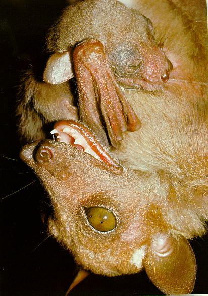
Echolocating and Non-Echolocating Bats
Echolocating bats have special external physical adaptations that allow them to reacquire the ecolocation pulse they send out. These adaptations are generally seen in the ears and face of echolocating bats. Notice that the ghostfaced bat has forward facing ears mounted low on its head and a highly sculpted face (i.e., it looks like it has been hit in the snout with a two-by-four). These facial ridges help deflect the returning echolocation pulse towards the ghost faced bat's ears. The fruit eating bat, by contrast, lacks these specializations. Notice the larger eyes and the unmodified snout. While this fruit eating bat does have fairly large ears they are not positioned well for the receiving of returning echos.
Echolocating Ghost Faced Bat
Non-Echolocating Fruit Eating Bat
Horseshoe bat
Long CF pulses
- Poor for target description
- Good for Doppler shift
Doppler Effect
Approaching sound: high frequency
Retreating sound: low frequency
Sound source & observer
Stationary and moving sounds
Doppler shifts measure velocity of targets
- Echo freqeuncy of insect wing beats
FM pulses good for
- Target description
- Range information
CF pulses good for
- Prey detection
- Increasing maximum range
Stages of Bat Flight
Search stage
- Species-specific sound pulse, 10 Hz
- Open space species
- Short CF pulses
- Long distance
- Closed space species
- FM pulse prominent
- Prey from background
- Eptesicus fuscus
- 2 cm sphere at 5 m
- 0.5 cm sphere at 3 m
Approach stage
- Turn head & ears towards target
- Increase pulse rate
- All species go to more FM pulses
- Make decision as to capture
Terminal stage
- Very high pulse rate
- FM pulses
- Multiple harmonics
Neurophysiology of Bat Auditory System
Typical mammalian ear
Auditory nerve to:
- Coclear nucleus (hindbrain)
- Inferior colliculus (midbrain)
- Auditory cortex (forebrain)
Specialisations
- Bat's hearing sensitive to frequency of its own sounds
- Hearing highly directional
- Higher levels of auditory pathway for pulse-echo delays
- Reduce sensitivity to other sounds (e.g., muscular contraction of middle ear prior to pulse; partially decouples inner ear from tympanum)
- Doppler shift neurophysiology
Maximally sensitive to a 60 degrees forward cone
Neural recovery of auditory system
Pulse return in 30 ms
Full recovery within 2 ms
Auditory cortex
- Systematic distribution of echo-ranging neurons
Return to 403 course page.